EN

动物肿瘤测量跟踪系统

品牌:比利时 Peira 型号:TM900 快速、精确测量动物肿瘤,肿瘤信息存档与跟踪!

产品介绍


品牌:比利时 Peira 型号:TM900

快速、精确测量动物肿瘤,肿瘤信息存档与跟踪!

产品特点:
手持式成像装置,实现立体成像
适合测量不同尺寸的肿瘤
方便使用:触屏式电脑,操作方便
内置软件,自动计算肿瘤尺寸,跟踪整个实验进展。


技术参数:
测量范围:0-25mm
最大肿瘤尺寸:
20mm*20mm*20mm
3D 测量精确度:<0.3mm
图像捕捉时间:0.1s
接口:USB 2.0
相机:1600*1200 像素(2MP)
工作距离:50mm


保证了高通量,精确、快速、可靠。全自动测量跟踪系统。实时数据分析与处理。


小鼠皮下注射肿瘤细胞,随后成瘤并逐渐长大。Peira TM900 肿瘤测量跟踪系统可以快速、精确测量动物肿瘤,完成肿瘤信息存档与跟踪。使用操纵非常简单,只需用扫描器对准肿瘤然后按下测量键,便可测量肿瘤体积。TM900 基于先进的立体成像技术,可以采集三维肿瘤图像。当肿瘤的结构光投射到扫描器的采集口时,系统根据结构光的变形系数自动计算出肿瘤的三维图像和体积。


“Caliper based tumor measurements can have issues with operator bias. Peira’s TM 900 contactless tumor volume measurement device is a faster and more accurate operator independent technology. The ability to electronically capture every raw image, together with a reference grid , coupled with easy transfer of raw data to standard data visualisation packages should offer major advantages over current methods.”
Unquote By : Timothy Perera, PhD Scientific Director and Janssen Fellow, Discovery Leader Long Tumor Stronghold Janssen Pharmaceutica


TM900 Management Interface

Save plot to file-可将统计图保存为PDF,BMP,PNG格式


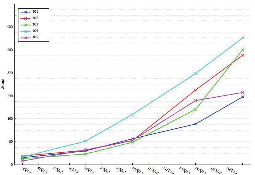
选择要测的动物-选择合适的探头-对准样本-按下开关-听到提示音松开开关得到真实照片和3D图像及数据(体积,体面积,高度)-选择是否接受当前测量-默认连测三次退出测量,得到平均值


TM900与手动测量具有显著的相关性,且偏差小于手动测量和人工测量对比优势:可视化、数据可追溯、数据直接读出、测量是三维测量。
目前谷歌学术文献45篇


Peira TM900 系统应用案例

SOX2 promotes lineage plasticity and antiandrogen resistance in TP53- and RB1-deficient prostate cancer


Ping Mu1, Zeda Zhang1,2, Matteo Benelli3, Wouter R. Karthaus1, Elizabeth Hoover1, Chi-Chao Chen4,5, John Wongvipat1, Sheng-Yu Ku6, Dong Gao1, Zhen Cao1,5, Neel Shah1,2, Elizabeth J. Adams1, Wassim Abida1, Phillip A. Watson1, Davide Prandi3, Chun-Hao Huang4,5, Elisa de Stanchina7, Scott W. Lowe4,5,8, Leigh Ellis6, Himisha Beltran9,10, Mark A. Rubin9,10, David W. Goodrich6, Francesca Demichelis3,9, Charles L. Sawyers


Science 06 Jan 2017:
Vol. 355, Issue 6320, pp. 84-88
DOI: 10.1126/science.aah4307


Sox2促进TP53和RB1缺失的前列腺癌的谱系可塑性和抗雄激素抗性
前列腺癌生长是由雄激素促发的。基于靶向雄激素受体(androgenreceptor,AR)的药物的雄激素剥夺治疗( androgen deprivation therapy, ADT)是治疗转移性前列腺癌病人的常用方法。虽然大多数病人会在开始治疗的时候产生应答,但是癌症几乎总会复发并且变得更具侵袭性,也更加致命。
一些癌症通过一种被称作细胞谱系可塑性的机制逃避靶向药物治疗,通过这种机制,肿瘤细胞获得一种存活不再依赖于药物靶标的细胞谱系的表型特征。

为了更加深入探究这种治疗抵抗性产生的机制,在一项新的研究中,来自美国斯隆凯特琳癌
症纪念中心的CharlesL.Sawyers及其团队利用体外和体内人前列腺癌模型证实这些肿瘤能够通过在表型.上由AR依赖性导管腔上皮细胞转化为不依赖于AR的基底样细胞,对抗雄激素药物恩杂鲁胺(enzalutamide)产生耐药性。这种细胞谱系可塑性是由TP53和RB1的功能性缺失引起的,并且受到重编程转录因子SOX2表达,上调的调节。

Sawyers团队进一步证实恢复TP53和RB1的功能或抑制SOX2表达能够逆转这种细胞谱系可塑性。
因此,肿瘤抑制基因发生的突变能够增加这种细胞可塑性,因此当遭受抗雄激素药物治疗时,这种细胞可塑性增加会通过细胞谱系转换促进前列腺癌产生抵抗性。
而在发表在同期Science期刊.上的另一项研究(Science, doi:10. 1126/science.aah4199)中,来自美国罗斯威尔帕克癌症研究所的LeighEllis和DavidGoodrich及其团队发现前列腺癌细胞是通过改变细胞谱系身份来逃避雄激素剥夺治疗的疗效,并且证实肿瘤抑制基因TP53和RB1的功能性缺失促进AR依赖性的导管腔.上皮细胞转化为不依赖于AR的基底样细胞,从而让前列腺癌对雄激素剥夺治疗产生抵抗性。

此外,研究人员借助临床前模型发现肿瘤抑制基因Rbl的缺失会诱导前列腺癌对治疗产生 适应性并促进前列腺癌转移。他们还发现另外一个基因EZH2表达增加也与前列腺癌对治疗 产生的适应性有关,或可用作治疗这种前列腺癌的靶点。利用EZH2抑制剂药物治疗产生抵 抗性的肿瘤可以增加前列腺癌对雄激素剥夺治疗的敏感性。
这些新发现为干扰甚至逆转这一致命过程提供了新的可能,同时也为预防和治疗这种致命性 的前列腺癌奠定基础。
该研究中采用比利时的Peira公司专利的TM900系统,对联合免疫缺陷小鼠(SCID mice) 体内异种移植实验过程中的肿瘤进行快速测量跟踪。


(C). Tumor growth curve of xenografted LNCaP/AR cells transduced with annotated hairpins. +Enz denotes enzalutamide treatment at 10 mg/kg orally 1 day after grafting. +Mock denotes DMSO treatment 

(C). Tumor growth curve of xenografted LNCaP/AR cells transduced with annotated hairpins.+Enz denotes enzalutamide treatment at 10 mg/kg orally 1 day after grafting. ....

....onceo tumors were palpable, tumor size was measured weekly by tumor measuring system Peira TM900 (Peira bvba, Belgium). All animal experiments were performed in compliance with the guidelines of the Research Animal Resource Center of the Memorial Sloan K ettering Cancer Center 


Fig. S3. Deletion of TP53 and RB1 using CRISPR-Cas9 system confers resistance to enzalutamide

Fig. S4. Murine organoids with Trp53 and Rb1 deletion lost epithelial features and gained resistance to enzalutamide


Figure I rhCNB inhibits the growth of tumor cells in vitro.Notes: (A and D) Cell viability was measured using MTT assays in SGC-7901, MGC -803, Bel-7 402 and HepG2 cells treated with the indicated concentrations of rhCNB for 24 h. (B and E) Cells were treated as in (A and D), and the degree to which rhCNB inhibited cell proliferation was measured using BrdU labeling. (C and F) SGC-7901 and Bel-7402 cells were treated with 3 mg/mL of rhCNB for 24 h, and cell proliferation was observed by microscope. Magnification: I0x * <0.05, *P <0.01 and ** <0.001. Abbreviations: rhCNB, recombinant human calcineurin B; DMSO, dimethylsulfoxide.